research-topics
- Simulated Atrial Fibrillation
- Investigation of Atrial Therapies
- Volume Conductor Model
- Ventricular Cancellation
Simulated Atrial Fibrillation
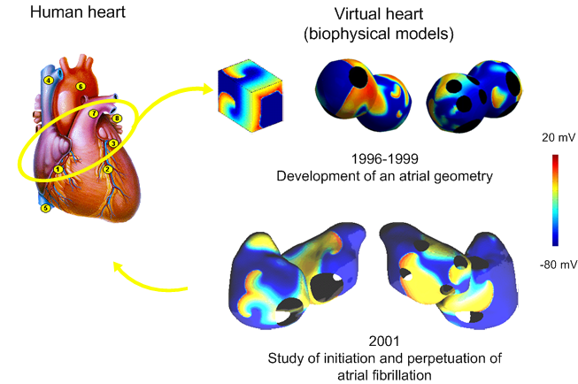

In 1996, the first model was developed. It was a simple cube into which the Beeler-Reuter membrane kinetics model was implemented. In 1999, the “peanuts model” was designed. It contained majors valves and veins. The membrane kinetics used was that of Luo-Rudy. In 2001, a more realistic biophysical model was developed based on MR images. The surface was meshed with triangular elements and more sophisticated membrane kinetics model was included (Courtemanche et al.). The biophysical model is currently used to study atrial fibrillation (AF) mechanisms: initiation and perpetuation.
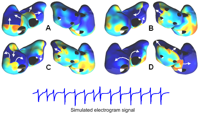
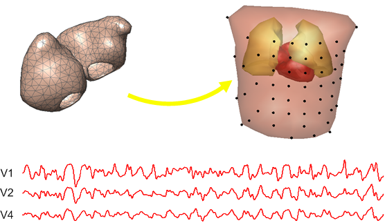
The biophysical model can simulate several types of arrhythmias, such as atrial flutter or atrial fibrillation. The figure above shows an example of sustained AF based on multiple reentries. The directions of the wavefront propagation are indicated by arrows. Furthermore, electrogram signals can be computed at any position on the surface of the atria.
Investigation of Atrial Therapies
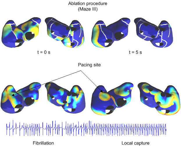
Two different types of therapies are currently investigated with the biophysical model: surgical ablation and therapeutic pacing. The result of simulated ablation lines confirmed the good performance of the Maze III procedure. Pacing demonstrated that atrial flutter can be pace-terminated and local capture can be reached in case of AF. An example of electrogram signal is displayed during AF pacing.
Volume Conductor Model
By extending the concept of the biophysical model to the entire thorax, the electric activity of the atria can be expressed as potential dynamics on the body surface. The lungs and the ventricular cavities are also included as well as their inhomogeneous conduction properties, so enabling the model to simulate realistic surface ECG signals reflecting pure atrial contributions.
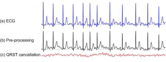
Ventricular Cancellation